Eye
Cherry Eye in Frenchies: What French Bulldog Owners Notice First
Published 2026-06-01; updated 2026-09-24. Educational resource from Los Angeles Bulldog Vet.
By Dr. Antonio Pedraza, DVM, MVM, cert SAS, MRCVS — Los Angeles Bulldog Vet at Sevilla Veterinary Hospital, Santa Monica.
How cherry eye appears in French Bulldogs and Frenchies, what to do in the early stage and what not to do, why the gland is put back rather than removed, and when a procedure is discussed.
- Cherry eye is a common name for prolapse of the third-eyelid tear gland.
- A pink mass at the inner corner is a reason for veterinary examination — not a home diagnosis of severity.
- Preserving the gland is usually preferred because it contributes to tear production.
- Surgery may be considered when prolapse persists or recurs; recurrence can still happen after repair.
- Sudden eye pain, cloudiness, or a protruding globe needs urgent care.
Cherry eye in a French Bulldog is a prolapsed third-eyelid tear gland: a smooth pink or red lump that appears at the inner corner of the eye, usually in a young dog and often overnight. It is rarely painful at first, it does not resolve reliably on its own, and it is treated by putting the gland back in place rather than removing it. This guide covers what Frenchie owners notice first, what to do and not do in the early stage, and when a procedure is discussed.
It is an educational article, not the procedural surgery page and not a substitute for an ophthalmic examination.
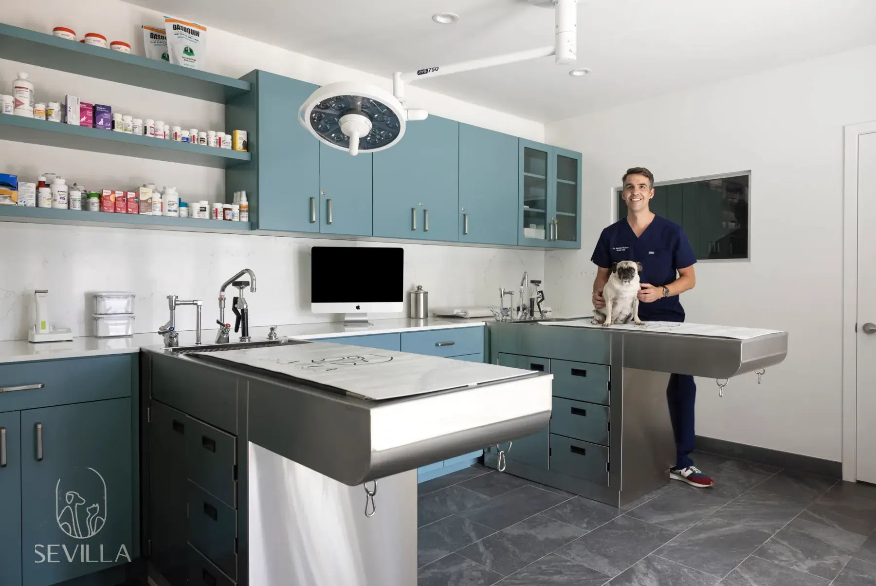
A clinician in navy scrubs stands behind a stainless exam table with a small light-colored dog facing the camera. Teal cabinetry and a ceiling surgical light are visible. No eye surgery is shown.
Why French Bulldogs are prone to it
The gland of the third eyelid (nictitans) is held in place by a small band of connective tissue. In French Bulldogs that attachment is often weak, and the breed combines a shallow eye socket, a prominent third eyelid and a habit of rubbing the face on furniture and carpets. When the gland swells even slightly, there is nowhere for it to go but out.
Because the weakness is anatomical, a Frenchie that has had cherry eye in one eye is at higher risk in the other. Both eyes are checked at every examination, and owners are asked to watch the second eye in the months after the first episode.
What Frenchie owners notice first
- A pink or red rounded lump at the inner corner of one eye, often noticed in the morning
- The lump seems to shrink or disappear for hours, then returns
- A wet face or brown tear staining below the affected eye
- Rubbing the face on the floor or pawing at the eye, with the dog otherwise cheerful
- In some dogs, the second eye following weeks or months later
Early stage cherry eye: what to do and not do
In the first days the gland is soft and only mildly inflamed, and this is the stage owners search about most. What helps: keep the eye clean with saline or a damp cloth, stop rough play and face-rubbing, use a harness rather than a collar, and book an examination in the coming days rather than weeks. Take a photograph and a short video for the history; a photograph helps the veterinarian, but it cannot confirm the diagnosis or how severe it is, and an online opinion is not a plan.
What does not help: massaging the gland back in as a long-term fix, over-the-counter redness drops, or waiting to see whether it settles. A gland that slips back briefly usually comes out again, because the tissue that held it has already stretched, and each week it spends exposed it becomes thicker and harder to reposition neatly.
What it is not
Cherry eye is not the same as entropion (an eyelid rolling inward), a corneal ulcer or an eye that bulges forward. Those look different, are more urgent in a flat-faced dog, and need prompt care. A Frenchie can have cherry eye and entropion at the same time, so a proper evaluation looks at the eyelids and the cornea as well as the gland.
Is my French Bulldog's eye popping out?
With cherry eye, the eyeball stays where it normally sits and a pink lump appears at the inner corner. An eye that is actually popping out is different: the eyeball itself is pushed forward out of the socket, often with the eyelids caught behind it. Veterinarians call this proptosis, and it is an emergency.
French Bulldogs are more prone to proptosis than most breeds because their eye sockets are shallow, so it can follow fairly minor trauma, rough play, or pressure on the neck. This is one more reason to walk a Frenchie on a harness rather than a collar. If it happens, keep the eye moist with sterile saline or a clean, wet cloth, stop the dog from pawing at it, do not try to push the eye back in, and go straight to the nearest emergency veterinary hospital, calling ahead so they are ready. Outcomes are better the sooner the eye is treated.
Why the gland is replaced, not removed
The nictitans gland produces a meaningful share of the tear film. Veterinary ophthalmology references recommend preserving and repositioning it whenever possible, because removal raises the later risk of dry eye (keratoconjunctivitis sicca), a condition that usually needs lifelong management.
Even after successful replacement, some dogs remain more prone to dry eye later, so tear testing and follow-up matter. Exact risk figures vary by source and population; your veterinarian will discuss what monitoring is appropriate for your dog.
What an evaluation may include
- Examination of both eyes, including the cornea and eyelids
- Tear production testing when appropriate
- Assessment for concurrent entropion, nasal fold irritation, or corneal disease
- Discussion of surgical timing, brachycephalic anesthesia considerations, and recurrence risk
When surgery may be discussed
Surgical replacement is discussed when the gland remains prolapsed or keeps returning. Timing depends on comfort, corneal health, age and overall anesthetic candidacy. Not every pink mark at the corner of the eye is cherry eye, and not every confirmed cherry eye is operated on the same day it is noticed.
No eye surgery is risk-free. Recurrence is possible after any technique. Outcomes depend on anatomy, surgical technique, aftercare and individual healing.
Urgent eye warning signs
Sudden squinting, acute eye pain, a cloudy cornea, or marked swelling should be treated as urgent. Do not wait for a routine cherry-eye consult if those signs appear. A visibly protruding eye is an emergency: go to the nearest emergency veterinary hospital, as described above.
Questions to ask about cherry eye
- Is this a prolapsed nictitans gland, or something else?
- What is the tear production status in each eye?
- What surgical technique are you considering, and what is the recurrence discussion for this dog?
- How will brachycephalic anesthesia and recovery be handled?
Local consultation for cherry eye
Stay on this page for early educational reading about signs, examination, and questions to ask. When you are ready for procedure-focused counseling in Los Angeles, continue to the dedicated cherry eye surgery page. For the wider map of Bulldog eye conditions, use the eye surgery hub. Los Angeles Bulldog Vet is the brachycephalic-focused practice of Dr. Antonio Pedraza at Sevilla Veterinary Hospital in Santa Monica, and evaluates candidates after records review and examination.
When to contact a veterinarian sooner
Sudden squinting, acute eye pain, a cloudy cornea, or a visibly protruding eye should be treated as urgent.
What to bring to a consultation
- Records from your primary veterinarian, including prior diagnoses and medications
- Recent lab work, imaging, or referral reports when available
- Short phone videos of breathing, sleep, eye changes, vomiting, or regurgitation if relevant
- A timeline of when signs started, what makes them worse, and what you have already tried
Examinations for Los Angeles Bulldog Vet patients take place at Santa Monica. You can request a consultation or use the referring-veterinarian pathway when another clinic is coordinating care.
Educational references
External sources are linked for further reading. We do not reproduce their text. Access date for link verification: 2026-07-24.
Educational information · Eye. This page does not replace an examination, diagnosis or case-specific treatment plan from a veterinarian.
Request a focused Bulldog consultation
Share records and concerns. Our team will follow up to schedule an examination in Santa Monica when appropriate.